

Computerized tomography (CT) what is it, what is used and how it is done
- 2704
- 167
- John Von
Computed tomography (CT), also known as computerized axial tomography (CT) or simply as "scanner", is a Image diagnostic technique used by X -ray technology To obtain detailed 3D images of the human body.
Content
Toggle- What is a computerized tomography (TAC)?
- How is a TAC?
- TACK TYPES: WITHOUT CONTRAST AND CONTRAST
- 1. CT without contrast
- 2. CT with contrast
- For what type of diagnoses or procedures is the TAC is used
- Bibliographic references
What is a computerized tomography (TAC)?
Since its invention in the 1970s, the CT has revolutionized medicine and has become an essential tool in the diagnosis and treatment of numerous diseases.
This diagnostic technique uses X -ray technology and a computer to generate three -dimensional and detailed images of a specific part of the body. Unlike conventional radiographs, which produce two -dimensional images, the TAC It allows a more precise and deep vision of organs, bones, tissues and blood vessels.
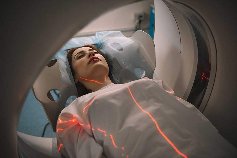
How is a TAC?
The CT machine consists of a large ring with a stretcher in the center where the patient is tende. When the tomography is performed, the machine sends x -ray beams through its body, which cross the tissues and are detected by sensors on the other side of the ring. The computer collects this information and creates images in "cuts" or "sliced" that doctors can see on a screen.
During the process, the stretcher on which the patient is lying slowly moves forward or backwards, allowing the machine to take images from different angles and levels. It is important that the examined person remains very still during the exam so that the images come out clear and precise.
The procedure It lasts approximately 10 and 30 minutes and is not painful. Once it ends, doctors analyze the images to diagnose diseases, identify injuries or plan treatments.
 What is love? And what is not
 TACK TYPES: WITHOUT CONTRAST AND CONTRAST
A CT without contrast and a tac with contrast are two types of computerized tomography that are used to obtain detailed images of the interior of the body. The main difference between the two is the use of a contrast medium in one of the procedures.
1. CT without contrast
In a tac without contrast, no contrast medium is used to improve the visualization of images. The CT machine takes X -ray images of the area of interest without any additional substance. These images They allow doctors to evaluate bone structure, masses and some other medical conditions.
A CT without contrast is useful in situations where the contrast is not necessary to obtain the required information or if the patient has any contraindication to receive the contrast, such as allergies or kidney problems.
2. CT with contrast
A CT with contrast implies the administration of a contrast medium to improve the visualization of certain structures and tissues inside the body. The contrast is a special substance that is introduced into the body orally, intravenously (injection into a vein) or enema. This substance makes the areas of interest more brilliant and in more detail in the tac images. The contrast is useful for better evaluating blood circulation, the presence of tumors, infections and other disorders which can be difficult to detect in a tac without contrast.
It is important to mention that, before administering the contrast, doctors should know the patient's medical history and verify whether there are associated contraindications or risks, such as allergies to the contrast substance, renal failure or heart problems.
Magnetic Resonance: What is and what are your applications
For what type of diagnoses or procedures is the TAC is used
Computed tomography (CT) is used to diagnose and evaluate a wide variety of medical conditions, as well as to plan therapeutic procedures. Some of the most common uses are the following:
- Trauma: with CT can identify bone fractures, internal hemorrhages, internal organ injuries and other damage in cases of accidents or serious injuries.
- Tumors: Tomography also allows you to detect tumors, evaluate its size, location and extension, and plan treatments such as surgery, radiotherapy or chemotherapy. It can be used to monitor the response to treatment and detect possible recurrences.
- Vascular diseases: Help visualize blood flow and detect obstructions, aneurysms, arteriovenous malformations and other anomalies in blood vessels.
- Infections and inflammatory diseases: This diagnostic test is capable of identifying abscesses, inflammation in organs, liquid collections and other alterations associated with infectious or inflammatory processes.
- Internal organ evaluation: It is useful for evaluating the state of organs such as brain, lungs, heart, liver, kidneys, pancreas and gastrointestinal system. Can detect abnormalities such as cysts, malformations, calculations and other medical conditions.
- Surgical planning: Provides detailed images of the patient's anatomy, allowing surgeons to precisely plan complex surgical procedures.
- Guided interventionist procedures: CT scan is also used to guide minimally invasive procedures such as biopsies, abscess drains or placement of medical devices (for example, stents).
- Neurological disease diagnosis: It is also used to evaluate patients with neurological symptoms, such as severe headaches, seizures or acute neurological deficits, since it can detect cerebral hemorrhages, brain tumors, cerebral infarctions, hydrocephalus and other conditions of the central central system.
These are just some examples of the many applications of computerized tomography in the diagnosis and treatment of medical conditions. As we can see, it is a valuable and versatile tool that continues to evolve and improve to provide more precise and effective medical care.
The electroencephalogram (EEG): a window to the brain
Bibliographic references
- Saba, l., Reason, e., and Suri, J. S. (2018). Multidetector computed tomography imageology: principles, head, neck and vascular systems. CRC Press.
- Radiologyinfo.Org (2021). Computed tomography (TC). Recovered from https: // www.Radiologyinfo.org/sp/info.CFM?pg = bodyct
- « Brain aneurysms, signs, diagnosis and treatment
- Graphic test of the house, what it is and how it is interpreted »