

Central Nervous System (CNS) Structure, Functions and Diseases
- 2649
- 545
- Lorenzo West I
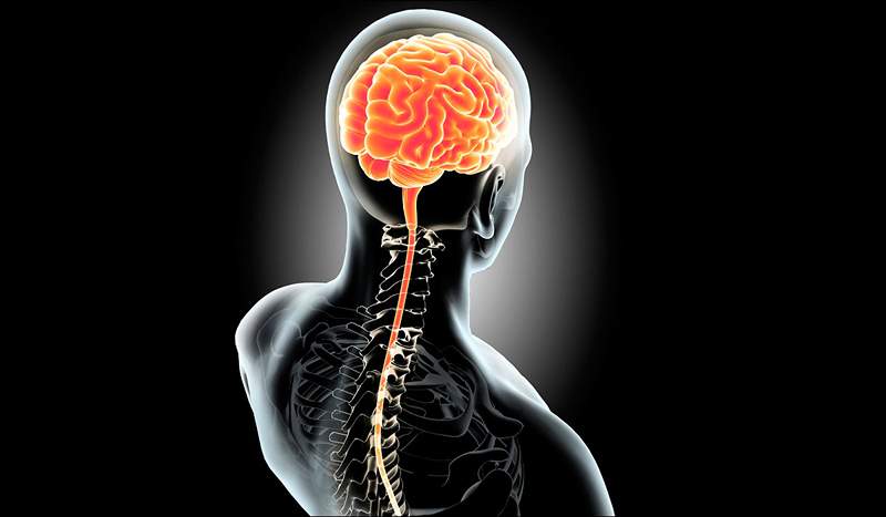
The central nervous system (SNC) is the part of the nervous system that is responsible for analyzing and integrating the information it receives from the internal and external environment, and of generating coordinated responses. It is formed by the brain and spinal cord. It is known as "central", since it integrates information from the whole body and coordinates the activity throughout the body.
The central nervous system has been studied for decades by doctors, anatomists and physiologists, but still keeps many secrets. Our thoughts, our movements, our emotions and our desires are generated inside, but we still have a lot to get to know all its mysteries.
Content
Toggle- ANATOMY OF THE CNC
- Differences between the central nervous system and the peripheral nervous system
- White matter and gray matter, main functions
- Glial cells
- Astrocytes
- Oligodendrocytes
- Microglias
- Spinal cord
- The cranial nerves
- The brain, anatomy and physiology
- Central nervous system diseases
- References
ANATOMY OF THE CNC
The central nervous system (CNS) is a complex structure that is found in the human body and is Responsible for controlling and coordinating most body functions. This system is made up of the brain and spinal cord, two organs that work together to transmit electrical and chemical signals throughout the body.
He brain It is the largest organ of the CNS and is protected by the skull. It is divided into two hemispheres that are connected by a structure called calloso body. Each hemisphere is composed of four lobes: frontal, parietal, occipital and temporal. Each lobe has specific functions, for example, the frontal lobe is involved in planning and decision -making, while the occipital lobe is involved in vision.
The spinal cord It is a nervous cord that extends from the base of the brain to the bottom of the spine. It is protected by a column of bones called vertebrae. The spinal cord is made up of nerve cells called neurons and support cells called glial cells. The spinal cord is responsible for transmitting nerve signals between the brain and the rest of the body. He is also responsible for performing autonomous functions, such as breathing and digestion.
The cerebral hemispheres
The neurons They are the cells that transmit electrical and chemical signals in the CNS. There are several types of neurons in the brain and spinal cord, each with specific functions. Sensory neurons are responsible for transmitting sensory information to the brain, while motor neurons are responsible for controlling muscle activity. Association neurons are responsible for communication between sensory and motor neurons, and are crucial for learning and memory.
In addition to neurons, there are several support cells in the CNS. Glial cells, also known as support cells, include astrocytes, oligodendrocytes and microglia. Astrocytes are glial cells that provide structural support and nutrition to neurons. Oligodendrocytes produce a substance called myelin, which helps transmit electrical signals more quickly along neurons. Microglia is a type of glial cell that has the defense function of the central nervous system.
Differences between the central nervous system and the peripheral nervous system
The nervous system is a complex network of tissues and organs that are responsible for coordinating and controlling body functions and responses to the stimuli of the environment. The nervous system is divided into two main parts: the central nervous system (CNS) and the peripheral nervous system (SNP). Although the two systems are intimately intertwined, there are large differences.
Within the main differences between The SNC and the SNP, is the difference in cell size. The axons of the nerves of the central nervous system (the thin projections of the nerve cells or neurons that transport the impulses) are significantly shorter. On the other hand, SNP nerves can be up to 1 m long (for example, the nerve that innervates the big toe), while in the CNS more time more than a few millimeters.
At a functional anatomical level, the Central Nervous System It is composed of the brain and spinal cord. The brain is the body's information control and processing center, while the spinal cord is responsible for transmitting information between the brain and the rest of the body. The CNS is responsible for vital functions such as thought, memory, emotions and movement.
He peripheral nervous system, On the other hand, it includes all the nerves that extend from the brain and spinal cord to the rest of the body. These nerves are divided into two main types: somatic nerves and autonomous nerves. Somatic nerves control voluntary muscles and body sensory information, while autonomous nerves control the involuntary functions of the body, such as breathing, heart rate and digestion.
Other Important difference between CNS and SNP is its capacity for regeneration. Much of the SNP has the ability to regenerate; If a nerve on a finger is cut, it can grow again. On the other hand, the CNS does not have this capacity.
 The sensory areas of the cerebral cortex
 White matter and gray matter, main functions
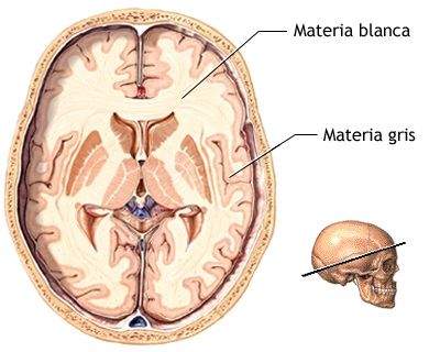
Gray and white matter are two main components of the central nervous system, which differ in its structure, function and location in the brain and spinal cord. The brain has a external cortex called gray matter and an internal zone that consists of extensions of white matter.
The Gray matter It is a layer of nerve tissue composed of neuronal bodies, dendrites and non -myelinized axons. It is mainly found in the cerebral cortex, in the basal nuclei and in the cerebellum. Gray matter is responsible for processing and transmitting sensory and motor information, as well as regulating cognitive and emotional functions.
On the other hand, the White matter It is a layer of nerve tissue composed of myelinized axons and glial cells. White matter is under the cerebral cortex and inside the brain and spinal cord. Its main function is to transmit nerve signals from one area of the brain to another and between spinal cord and brain.
The White matter The brain has always been considered as a passive support of neuronal activity. Its main function is the transmission of brain information. This substance moves the electrochemical pulses emitted by the brain to the rest of the body. Its main function is to coordinate communication between the different systems of the human body, both inside and outside the brain. Recent research shows that also intervenes in learning, cognitive and emotional processing, and in the generation of mental illnesses.
The Gray matter Lack of myelin, he is not able to quickly transmit nerve impulses. On the other hand, its function is related to information processing and therefore also of reasoning. Is responsible for elaborating the appropriate responses to the different stimuli.
The white substance and gray substance of the brain: function and comparative
Glial cells
Glial or neuroglia cells are a non -neuronal cell type that plays a fundamental role in the central nervous system. They are often called "support cells" because they support and nourish neurons, but they are also responsible for a variety of important functions, from brain formation to protection against damage and tissue repair.
Among its main functions is to control the cell ionic microenvironment, neurotransmitters levels and the supply of cytokines and other growth factors.
Without glial cells, the developing nerves would be unable to reach their destinations and, if they do not find their way, they are not able to form functional synapses.
There are three main types of glial cells: Astrocytes, oligodendrocytes and microglia. Each of them has unique functions and characteristics.
Astrocytes
Astrocytes are more common glial cells and are found throughout the brain. They provide physical and metabolic support to neurons, making sure they are well nourished and protected. Astrocytes also play an important role in the formation of the hematoencephalic barrier, a protective barrier that separates the blood from the brain to avoid the entry of harmful substances.
These cells have numerous projections and supply the blood to anchor neurons. They also regulate the local environment by eliminating excess ions and the recycling of neurotransmitters. Astrocytes are also divided into two different groups: protoplasmic and fibrous.
Oligodendrocytes
Oligodendrocytes are glial cells that produce myelin, a substance that covers the neurons axons. Myelin acts as an electric insulator, which accelerates the transmission speed of nerve signals and allows them to send signals quickly and efficiently ... the lack of myelin or the degeneration of oligodendrocytes can lead to diseases such as multiple sclerosis.
Microglias
The microglia are specialized immune cells that are found in the brain and spinal cord, forming part of the central nervous system. They are the smallest glial cell type and represent between 5% and 20% of all brain cells.
Microglia have a fundamental role in the defense and protection of the central nervous system, since are responsible for detecting, eliminating and degrading pathogens, dead or damaged cells and other waste cell phones that can endanger brain health.
In addition, microglia also have an important role in the modulation of the inflammatory response and in the regulation of synaptic plasticity and neurogenesis, which gives them a key function in cerebral homeostasis and in the adaptation of the brain to the different situations and stimuli.
Spinal cord
The spinal cord is a nervous structure that is found within the vertebral channel, inside the spine, and extends from the brain trunk to the lumbar region. It is part of the central nervous system and has a cylindrical and elongated shape.
The spinal cord is a communication channel that connects the brain with the rest of the body, transmitting nerve impulses from the brain to the muscles and organs, and vice versa. It is formed by a set of neurons and nerve fibers that are organized in different structures and nerve pathways, and that are responsible for the transmission of sensory, motor and autonomous information.
In addition to its information transmission function, the spinal cord also plays an important role in the integration of nerve impulses, the coordination of movements and the regulation of autonomous functions, such as breathing, heart rate and digestion. That is why any injury or damage to the spinal cord can produce important disorders and disabilities in body functioning.
Through the spinal cord you can coordinate the movement of the muscles throughout the body.
The spinal cord travels the back of the organism and carries the information between the brain and the body, but also carries out other tasks. From the brain trunk, where the spinal cord is found with the brain, there are up to 31 spinal nerves connected to the nerves of the SNP, which are responsible for giving sensitivity and function to the skin, muscles and joints.
Motor orders travel from the brain, pass through the spine and reach the musculature. Sensory information travels from sensory tissues (such as skin) to the spinal cord and, finally, to the brain.
The spinal cord contains special circuits for reflex responses, such as the involuntary movement that a hand could do if the finger comes into contact with a flame.
Circuits within the spine can also generate more complex movements such as walking. Even without brain participation, spinal nerves can coordinate all the necessary muscles to walk. What they cannot do will be to initiate, stop or make changes in said movement, because this is the exclusive function of the brain.
 Sensitivity to physical sensations: hyperesthesia
 The cranial nerves
Have 12 pairs of cranial nerves that arise directly from the brain and pass through holes in the skull to travel along the spinal cord. These nerves collect and send information between the brain and the different parts of the body, especially the neck and head.
Of these 12 pairs, the olfactory nerve, the optic and the terminal nerves arise from the anterior brain and are considered to be part of the central nervous system:
- Olfactory nerves: They transmit information from the smell of the upper section of the nasal cavity to the olfactory bulbs at the base of the brain.
- The optical nerves: They carry the visual information from the retina to the primary visual nuclei of the brain. Each optical nerve consists of around 1.7 million nerve fibers.
- Terminal cranial nerves: They are the smallest of the cranial nerves, their role is not yet clear. Some believe that they can be vestigial (an evolutionary byproduct that has no remaining function) or that participate in the function of pheromones (secreted hormones sensors that cause answers in social animals).
The brain, anatomy and physiology
The brain is the most complex organ in the human body. The cerebral cortex (the outermost part of the brain and the largest part in volume) contains between 15-33 million neurons, each of which are connected to thousands of other neurons. In total there are about 100 billion neurons and 1.000 of glial cells that constitute the human brain.
The brain is the central control module of the body and coordinates a multitude of tasks. From the physical movement to the secretion of hormones, through the creation of memories and the feeling of emotion, among many others.
To carry out all these functions, some sections of the brain have specific functions. However, many of the higher functions such as reasoning, problem solving or creativity, involve different areas that work together in network.
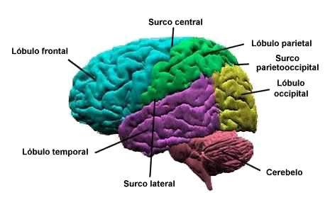
The brain is divided broadly in four lobes:
- Temporary lobe: The temporal lobe is important for the processing of sensory and emotional information. It also participates in the Fixing long -term memories in relation to the hippocampus. Some aspects of language perception are also found here.
- Occipital lobe: The occipital lobe is the visual processing region of the mammals' brain. Primary visual cortex damage can cause blindness.
- Parietal lobe: The parietal lobe integrates the sensory information that includes touch, spatial perception and orientation. Tactile skin stimulation is ultimately sent to the parietal lobe. He also plays a role in language processing.
- Frontal lobe: Located on the front of the brain, the frontal lobe contains most dopamine neurons and is involved in attention, reward, short -term memory, motivation and planning.
The following are some specific regions of the brain with a summary of their functions:
- Basal ganglia: Basal nodes are involved in the control of voluntary motors and the learning process. The diseases that affect this area are Parkinson's disease and Huntington's disease
- Cerebellum: It is mainly responsible for the control of fine and precise movements, it also participates in the language and attention process. If the cerebellum is damaged, the main symptom is the interruption of motor control, known as the ataxia.
- The Broca area: This small area located on the left side of the brain (sometimes right in left -handed people), has an important function in language processing. When the person is damaged, he presents difficulties in speaking, but he is still able to understand speech. Stuttering is sometimes associated with low activity in the Broca area.
- Hard body: It is a wide band of nerve fibers that unite the left and right hemispheres. It is the largest structure of white matter in the brain and allows the two hemispheres to communicate. It has been seen that dyslexic children have the smallest callosum, while left -handed people, ambidiestras and musicians, usually have it larger.
- Spinal bulb: It is under the skull, it is an essential structure for numerous involuntary functions, such as breathing, sneezing, vomiting and maintenance of correct blood pressure.
- Hypothalamus: It is just above the brain trunk and has the approximate size of an almond. Segregates a whole series of neurohormones and influences a variety of answers including body temperature control, hunger and thirst.
- Tálamo: placed in the center in the brain, the thalamus receives sensory and motor entries and transmits them to the rest of the cerebral cortex. It is involved in the regulation of consciousness, sleep and alertness.
- Amygdala: They are two almond -shaped nuclei in the internal zone of the temporal lobe. They are involved in decision -making, memory and emotional responses, especially negative emotions.
Central nervous system diseases
A system as as complex and extensive as the CNS can work badly for enough reasons. Below are the main causes of the disorders that affect the central nervous system:
The CNS is susceptible to many diseases and injuries, ranging from infection to cancer.
- Trauma: Any significant lesion in the brain or spinal cord can cause negative health consequences. Depending on the site of the lesion, the symptoms can vary widely, from motor paralysis to cognitive or humorous disorders.
- Infections: various microorganisms and viruses can invade the central nervous system. These include fungi (cryptococcal meningitis), protozoa (malaria) bacteria (leprosy) and virus of different types.
- Degeneration: spinal cord or brain can degenerate, causing different problems depending on what areas are degenerate. An example is Parkinson's disease, which implies the gradual degeneration of dopamine producing cells in the black substance of basal ganglia.
- Structural defects: The most common examples within this category are birth defects; An example is anencephaly, where the main parts of the skull, brain and scalp are missing at birth.
- Tumors: both cancerous and non -cancerous tumors can affect parts of the central nervous system. Both types can cause damage and produce a series of symptoms, depending on where they develop.
- Autoimmune disorders: In some cases, an individual's immune system can attack healthy cells. For example, acute disseminated encephalyitis is characterized by an immune response against brain and spinal cord, attacking myelin (nerve insulation) and, therefore, destroying white matter.
- Cerebral vascular accident (AVC): A stroke is an interruption of the blood supply to the brain; The consequent lack of oxygen makes the tissue of the affected area die.
References
- Bradford, h.F. (1988). Fundamentals of neurochemistry. Barcelona: Labor.
- Carpenter, m.B. (1994). Neuroanatomy. Fundamentals. Buenos Aires: Panamerican Editorial.
- Delgado, j.M.; Ferrús, a.; Mora, f.; Rubia, f.J. (EDS) (1998). Neuroscience Manual. Madrid: Synthesis.
- Diamond, m.C.; Scheibel, a.B. And Elson, L.M. (nineteen ninety six). The human brain. Work book. Barcelona: Ariel.
- Guyton, a.C. (1994) Anatomy and Physiology of the Nervous System. Basic neuroscience. Madrid: Pan American Medical Editorial.
- Kandel, e.R.; Shwartz, J.H. And Jesell, T.M. (eds) (1997) Neuroscience and behavior. Madrid: Prentice Hall.
- Martin, J.H. (1998) Neuroanatomy. Madrid: Prentice Hall.
- Nolte, J. (1994) The human brain: Introduction to functional anatomy. Madrid: Mosby-Doyma.