

Cranial nerves, what are your functions?

- 2315
- 41
- Jeffery Jones
If we say the name of hypogloso nerve, perhaps many do not sound them. However, if we talk about the vagus or trigeminal nerve, they may be better known. The three nerves belong to the twelve cranial nerves, also known as the twelve cranials. The name of cranial pares.
Content
Toggle- Where are the cranial nerves?
- Cranial nerves function
- Olfactory (i)
- Optic (II)
- Oculomotor (III) and Troclear (IV)
- Trigeminal (v)
- Abduens or external eye motor (VI)
- Facial (VII)
- Vestibulomotor or auditory (VIII)
- Glosopharyngeal (IX)
- Vago (x)
- Accessory (XI)
- Hypogloso (XII)
- Bibliography
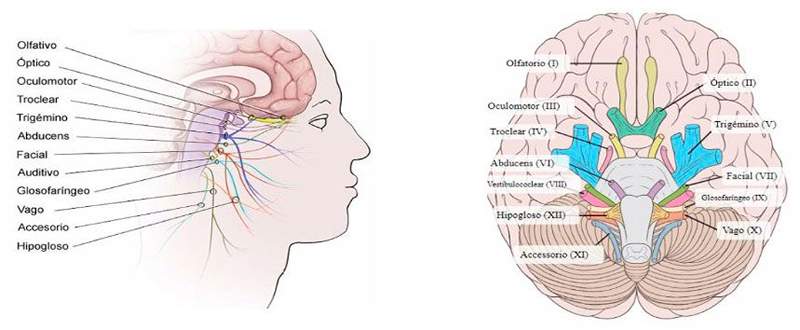
Where are the cranial nerves?
Most of them are located in the brain trunk, specifically in the midbrain, the bridge and in the spinal bulb. The first two cranial nerves are the only ones that are located outside the brain trunk. Specifically, the olfactory nerve (i) and the optical nerve (II).
The Oculomotor (III) and the Trocreal (IV) nerves, leave from the midbrain. On the bridge are the trigeminal (V), the abducens or external ocular motor (VI), the facial (VII) and the viewubulococcar or auditory (VIII). Finally, the glossopharyngeal nerve (IX), the vagus (x), the accessory (XI) and the hypoglossus (XII), are found in the spinal bulb.
Cranial nerves function
The cranial nerves have a motor and sensory and mixed function. Some of them belong to the somatic nervous system and others to the autonomic nervous system. On the one hand, several of them transport sensory information to the central nervous system (sensory function), and others do so in the opposite direction, from the CNS to different areas of the body (motor function). Others combine both functions, both motor and sensory.
Olfactory (i)
Its function is sensory. Its axons end in the olfactory bulb. It is composed of axons of neurons whose dendrites and cell bodies are in the olfactory mucosa.
Optic (II)
The optical nerve is formed by retinal cell axons that They transport visual information from the eyes to the brain. The two optical nerves converge in optical chiasma. At this point, some fibers of each nerve cross on the opposite side. From here, through the optical tract, visual information is transported to the thalamus. The function of this nerve is sensory.
Oculomotor (III) and Troclear (IV)
Its functions are motor. They act on the musculature of the eye. The axons start from the midbrain and have fibers of the autonomic nervous system that innervate the smooth muscles of the eyeball, specifically, the constrictor muscles of the iris and the ciliary musculature that is responsible for the control of the form of the iris.
Trigeminal (v)
This nerve if it places on the bridge. It is a motor and sensory nerve. It has three sensory branches that facilitate sensory information from different points of the face, tongue and mouth. Motor fibers innervate the muscles of the jaw that control chewing.
Abduens or external eye motor (VI)
It is a motor muscle that Control the straight muscle of the eye. When activated, it is accompanied in a coordinated way with the oculomotor and troclery nerves.
Facial (VII)
The facial nerve is mixed. On the one hand, It has a somatic motor component that inverts the muscles in charge of facial expression. Also invert autonomic nervous system fibers that are directed towards the tear and salivary glands. On the other hand, the sensory fibers take information from the gustatory papillae of the previous part of the language and are involved in the sense of taste.
Vestibulomotor or auditory (VIII)
Sensory nerve with two differentiated branches. The cochlear branch innervates the cochlea, inserted in the audition body. The vestibular branch transports information from the vestibular device (equilibrium organ).
Glosopharyngeal (IX)
Located in the spinal bulb. It is a sensory and motor nerve with Somatic and visceral components. The main function of the glossopharyngeal nerve is the collection of information from the mucous membranes of the pharyngeal region and the posterior third of the tongue. Somatic motor innervation is carried out in the striated muscles of the pharynx and visceral on the parotid gland.
Vago (x)
It reaches head of the head such as the pharynx, larynx and trachea; and to trunk structures such as heart, lungs and digestive system. It is a sensory and motor nerve that intervenes in a large number of somatic and visceral functions. On the one hand, it reaches the striated musculature of the palate, the pharynx and the larynx in order to control the swallowing. It also collects sensory information from a large part of the viscera of the abdomen and thorax. His role on the autonomic nervous system stands out in aspects such as heart rate, gastric secretion and intestinal peristalism.
Accessory (XI)
It is a motor nerve. It is considered an accessory nerve of the vagus nerve since their fibers pass through branches of the latter such as those of the pharynx and larynx, thoracic and abdominal viscera. On the other hand, he also reaches his shoulder and neck to control his movements.
Hypogloso (XII)
It is a motor nerve that controls the Language musculature.
Bibliography
April, a., Ambriosio, e., Rosario de Blas, M., Caminero, a., Garcia, c., Pablo, J. and Sandoval, and. (2005). Biological basis of behavior. Madrid: Sanz and Torres.