

Morphogenesis of the nervous system how the brain forms
- 3498
- 251
- Miss Drew Stroman
Morphogenesis is the process by which structures and bodily patterns are formed during the development of an organism. This process implies a series of cell and molecular events that lead to cell differentiation, tissue and organ organization, and the formation of growth and differentiation patterns. Morphogenesis is essential for the normal development of organisms, from unicellular organisms to the most complex multicellular organisms.
In animals, morphogenesis is particularly important for the formation of body structure, organ formation and cellular specialization during fetal development. In plants, morphogenesis is important for the formation of structures such as leaves, stems, flowers and roots.
Content
Toggle- Cell reproduction
- Fertilization
- Neural tube formation
- References
Cell reproduction
Cell reproduction is the process by which a cell is divided into two or more daughter cells, each with an identical copy of the genetic material of the stem cell. This process is essential for the growth, development and repair of organism's fabrics, as well as for sexual reproduction in multicellular organisms.
There are two main types of cell reproduction: Mitosis and meiosis. Mitosis is the process of cell division that produces daughter cells identical to the stem cell, and is essential for the growth and repair of body tissues. Meiosis, on the other hand, is the cell division process that produces daughter cells with half the number of chromosomes of the stem cell, and is essential for sexual reproduction, since it produces sex cells (gametes) that merge to form to form A new organism.
As we said, the cells that reproduce sexually do so through the Meiosis, The nuclear division process by which sex cells (gametes) are formed. Each chromosome from a cell is divided into two (diploid). During meiosis, these diploid chromosomes pairs double and separate so that each meiotic sex cell has only one chromosome (haploid) of each pair. Two successive meiotic divisions result.
During the life cycle of Organisms that reproduce sexually, fertilization results in the fusion of haploid gametes (sperm and ovules) that produce zygote. Divided by asexual cell reproduction, the zygote suffers from cell differentiation, so the cells become structural, functionally and biochemically different from each other.
Fertilization
Human fertilization is produced by the penetration of a male sperm in a female ovule. The result of fertilization is a cell (zygote) capable of producing cell division to form a new individual.
The zygote will begin to be divided by successive mitosis and will pass through the following phases:
- Morula: From twelve to sixteen homogeneous cells (third day).
- Blastula: As the cell division continues, an interior space is formed in the morula. The blastula is implanted in the uterus at the end of the first week after fertilization.
At this time, the embryo is an album consisting of two layers of cells, a superior (epiblast) and a lower (hypoblast). In the third week the gastration phase begins that begins by the training in the epiblast of the primitive line.
The primitive line is a small invagination of the epiblast produced by the migration of cells of this layer towards an intermediate position between the epiblast and the hypoblast. As a result of this migration, a third layer of the embryo is formed, the mesoderm, which is between the ectoderm (old epiblast) and the endoderm (old hypoblast).
From these three layers, ectoderm, mesoderm and endoderm, all the tissues and organs of the new individual will be formed.
- Of the Ectoderm They come skin, hair, nails and SN (neurons and glial cells).
- Of the endoderm Visceral organs come (digestive and respiratory system).
- Of the mesoderm They come muscles and bones. It also contributes to the formation of the SN.
Neural tube formation
Shortly after its training, the Ectodermo thickens through the midline where the neural plate arises (Day 18-20). As the growing neural plate bends on the sides forming the neural channel. This neural channel is completely closing the neural tube.
Around twenty -three embryonic life, the neural tube It is practically closed, except at the ends, where we find the Neuroporo Rostral and the flow. If the closure of these neuroporo is not done correctly, there are a wide variety of congenital malformations. For example, when the error takes place in the closure of the caudal neuroporo, malformations occur in the spinal cord, such as the Bifida spine. If the error occurs in the Rostral Neuroporo, they occur Malformations in the brain and skull, that is split.
A part of the embryonic cells is outside the neural tube when the neural ridges are closed and forms. From these neural ridges they will be derived from the SNA, the SNP sensory neurons, the SNP glia and the meninges. That is, from the neural ridges will derive the neurons that have their cell body outside the CNS, in the peripheral ganglia.
In this period of development, The neural tube is formed by a layer of tissue called neuroepitelium, that has a characteristic structure. The neuroepitelium is formed by germ cells that are distributed between the ventricular and the marginal area. This distribution gives the neuroepitelium a pseudoestratified appearance that gives the impression of being formed by layers. When the neural tube is closed, neuroepithelium cells begin to divide by mitosis, and the cells that have finished their mitotic period in the ventricular zone are between it and the marginal, and configure the intermediate zone.
The anterior part of the tube will form the brain and the rest will form the spinal cord.
The neural tube cavity will lead to the ventricular SNC system.
At the end of the fourth week in the neural tube, three vesicles are distinguished in the cephalic region:
- Prosencéfalo
- Midbrain
- Romboenfalo
- Flow region of the neural tube.
In the fifth week, it is possible to distinguish five brain vesicles. Prosencephal. The rhomboenphal is also divided into two vesicles: metencephalon and mylencephalon. The mesencephalic gallbladder is not divided.
From the fifth week in the neural tube, five vesicles are distinguished in the cephalic region:
- Telencéfalo
- DIENTINFALO
- Midbrain
- Metencephalon
- Mielencephalon
- Flow prolongation of the neural tube.
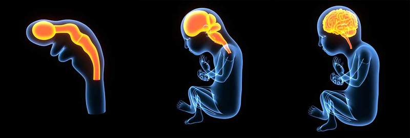
In the course of development, the accelerated mitotic process experienced by cells gradually change the structure of the neuroepitelium. This process does not take place in a homogeneous way, but there is a differential growth of the neuroepitelium on the wall of vesicles, which causes the appearance of the different structures of these divisions. For example, The hemispheres will end up surrounding most of the diencephalon and the midbrain. It is precisely this great development of Telencéfalo that responsible for the superior capacities of the human nervous system. When the development ends, 70% of the neurons that form the brain will be found in the cerebral cortex.
References
- Gilbert, s. F. (2005). Developmental biology. 7a. ed. Mexico: Panamericana.
- Sunderland, Mary and., Morphogenesis. Embryo Project Encyclopedia (2008-05-09). ISSN: 1940-5030
- University of Salamanca. Definition of Concepción and its relationship with fertilization. Medical-Biological, Historical and Etymological Dictionary.
- « Serial murderers How does a normal person become a serial killer?
- Pere Bonet, elected new president of the Catalonia mental health cluster »