

The septal area, anatomy and function

- 2613
- 574
- Ryan Bogisich
Thanks to the advances in neuroscience and anatomy, today we know an area in the brain located in the frontal lobe whose diverse connections along the rest of the brain make it a key area to carry out different emotional and regulation functions, among many others. Today we talk about the characteristics of the septal area and of the Septal nuclei, regions that are part of the limbic system and whose characteristics are detailed below.
Content
Toggle- What is the septal area?
- The septal nuclei
- Septal Area Functions
- Septal area injuries
- Bibliographic references
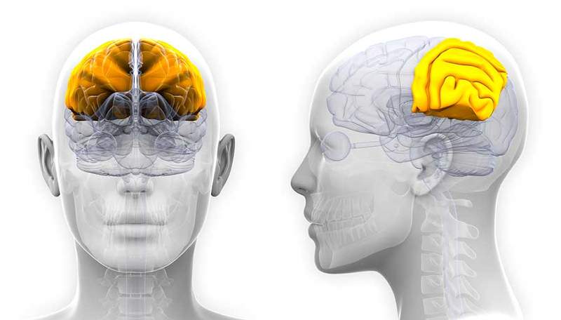
What is the septal area?
He septal area It is an area of the brain that is located in the inferosterior area of the medial face of the frontal lobe, located in front of the terminal sheet, a sheet composed of gray matter that connects the optical chiasma and the previous corner. It is also next to the lower area of the corpus callosum. A structure composed of nerve fibers that is responsible for unifying the two cerebral hemispheres.
The septal area achieves regulate emotional state Thanks to its ability toniby other brain areas responsible for the emotional and stressor process, such as the amygdala, a key region whose function is very importantly associated with the reaction of fear and emotions, among others. In addition, this area is involved, while many other structures, in emotional and pleasure processing.

The septal nuclei
Within the septal area are the Septal nuclei, a group of cell nuclei that are in front of the corpus calloso and adjacent to the septum. They are considered part of the limbic system and connect with other structures of the same system.
Among the functions of the septal nuclei, there is their participation in emotional, sexual, aggressive behaviors, as well as a modulation of autonomous functions and certain implications in attention and memory through cholinergic neurons.

The septal nuclei receive adhesions (neurons that arrive) from the hippocampus, a structure considered key in the memory processes. They also receive adhesions from the tonsil, the ventral tegmental area and several hypothalamic nuclei, among other structures.
In turn, the septal nuclei are directed towards the hippocampus and the dentated turn, a brain area also associated with memory and memories.
 The parietal lobe: Anatomy and function
 Septal Area Functions
As we have previously indicated, the septal area and in turn the septal nuclei have close connections with the hippocampus and the medial hypothalamus, very important structures in the limbic system that are in functions key such as emotions, memory, organic balance or level alert. The septal area seems to produce an inhibition in the structures of the limbic system getting decrease alert level. This provides improvements in memory and attention that are not diminished by a high state of stress. Thus, the septal nuclei manages to modulate the pleasant sensations and the extreme states of alert, managing to integrate the different cognitive processes such as motivation, emotion or memory to achieve a state of stillness and balance.

In addition, it has been proven as the stimulation of the septal nuclei through electrodes, they lead to intense pleasure responses that make the body seek pleasure again and again despite achieving later negative consequences.
Septal area injuries
The lesions in the septal area or in the septal nuclei have shown certain deficits in different fields of human and animal behavior. When damage occurs in these areas, Highly reactive behaviors may occur to sexual or food stimuli. This is because there is a lack of regulation in the limbic system due to the cessation of the inhibition that this region processes in its normal operation.
In addition, the connections that this area maintains with structures such as the hippocampus make memory or learning can also be harmed. As we have seen before, when this area is stimulated, the pleasant responses are very important. This has been shown even in studies with animals whose septal areas have been stimulated. After stimulation, animals have maintained obsessive pleasant responses even if they were followed by an aversive consequence. That is why the importance that this brain area has in the feeling of pleasure is confirmed.

Bibliographic references
- González-Pérez, m. N., & Olvera-Cortés, M. AND. (2017). The septal area: anatomy, physiology and its relationship with memory. Mexican Neuroscience Magazine, 18 (2), 32-41.
- Valdés-Sosa, m., & Lage, to. (1991). The septal area: an overview. Neurology Magazine, 7 (2), 47-54.
- Gómez-Ramírez, J., & Sanz-Martin, to. (2008). The septal area: anatomy, physiology and its relationship with neuropsychiatric disorders. Neurology Magazine, 47 (10), 546-554.
- Salgado-Pineda, p., & Delgado-García, J. M. (1993). The septal area: anatomy, physiology and functions. Neurology Magazine, 21 (121), 224-233.
- González-Pérez, m. N., & Olvera-Cortés, M. AND. (2018). The septal area and its relationship with emotion and social behavior. Neurology Magazine, 67 (5), 183-190.
- Hernández-González, m., & Sánchez-Sánchez, R. (2017). The septal area: anatomy, neurochemistry and its relationship with anxiety. Magazine of Psychiatry and Mental Health, 10 (3), 153-162.
- Autonomic-Hypothalamic-Limbic Systems. David l. Felten . 2016. https: // www.Scientedirect.com/science/article/pii/b9780323265119000163
- Septal Area. Dr Craig Hacking. https: // radiopaedia.org/articles/septal-area