

Glial cells what are, types and functions
- 2506
- 146
- Hugh Greenholt
When talking about the brain, you begin to think about neurons and their communication networks immediately. It is true, the brain and the nervous system work because neurons talk to each other, they touch each other exchanging information and playing the role of director in the comedy of life. ¿But what would happen if the glia was missing? In this Psychology-online article, we will talk about the glial cells, To better understand What are, the different types and their functions. Let's look at the definition, classification and characteristics of glial cells.
You may also be interested: White brain substance: definition, structure, functions and index injuries- What are glial cells
- Glial cell types
- GLIAL CELL FUNCTIONS
- Structure of glial cells
- Differences between neurons and glial cells
What are glial cells
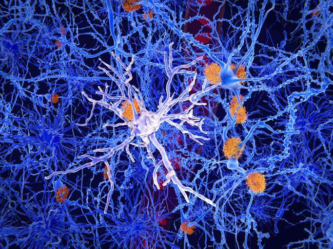
Gly, or neuroglia, is the most important cellular component of the nervous system, being 10 to 50 times larger than neurons. The name was introduced in the mid -nineteenth century to indicate The amorphous substance or "nervous cement" that surrounded and sustained neurons. Subsequently, the cell nature of the barrier and the existence of different specialized types was recognized.
By definition, glial cells are A type of cells of the nervous system which compose, together with the neurons, the central and peripheral nervous system. Described for the first time by the German physiology Rudolf Virchow in 1860, they carry out many auxiliary activities to the operation and survival of neurons, offering them above all a mechanical and nutritious support and exercising a metabolic control of Milieu intercellular.
Glial cell types
Like we can differentiate between various types of neurons, we can also find types of glial cells. In the nervous system of vertebrates there are two different types in terms of size and embryonic origin:
Macroglia
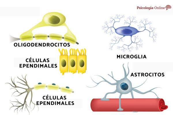
The classification of glial cells begins with 3 large types: macroglyia and microglia. The macroglyia, greater and of neuroectodermal origin, which in the central nervous system (CNS) includes Astrocytes, oligodendrocytes and ependymal cells. The latter are the cellular elements that cover the brain ventricles and the Central Medulla.
In the peripheral nervous system (SNP), the macroglia is represented by the Schwann cells, equivalent to the oligodendrocytes of the central and responsible for the formation of myelinic pods of peripheral axons and satellite cells, which delimit the outer surface of neurons in the spinal ganglia.
Microglia
Microglia, with smaller cells of mesodermic origin, differs from macroglyia in size and origin, and represents the population of Immunocompetent cells of the central nervous system. The latter have a function similar to that of macrophages and, through interleucin secretion, can influence not only immune response at the nervous system level, but also in neuronal activity and reactivity. Microglia represents approximately 10% of nervous system cells and, although present throughout the cerebral parenchyma, has a variable density from region to region, reaching the highest concentrations in the hippocampus, in the basal ganglia and in the black substance.
GLIAL CELL FUNCTIONS
Gly cells, in addition to providing support to neurons, They control the internal brain environment, They participate in the formation of specialized structures such as the hematoencephalic barrier and myelin sheath, ensure the isolation of nerve cells and their protection against strange agents or traumas. Other functions performed by glial cells are:
- Collect neurotransmitters molecules of the extracellular fluid (many neuroglia are equipped with neurotransmitter receptors).
- Compose the blood brain barrier that actively controls the passage of nutrients and other molecules of the blood torrent to neurons and vice versa.
- During neurobiological development, some specialized glial cells They guide neurons migration immature towards the appropriate places of the brain where they will be developed, and support the prolongation of axons towards their target cells.
The fundamental role of glia in the development and functioning of the nervous system is also reflected in its participation in many important neuropathologies. Neuroglia are a source of numerous tumors (called gliomas) of the brain, retina or spinal cord; In fact, in the brain of the adult animal, they face continuous mitotic divisions, which increases the probability that an evil mutation responsible for uncontrolled proliferation will occur.
After seeing the general functions of glial cells, let's see what each type of glia cells is for:
Satellite cells (SNP)
- They surround cell bodies in the ganglia.
- They regulate oxygen levels and carbon dioxide, nutrients and neurotransmitters around ganglion neurons.
Shwann cells (SNP)
- They wrap the axons in the peripheral nervous system
- Responsible for the myelination of peripheral axons
- Participate in damage repair processes
Ependymal cells (CNS)
- They upholster the encephalic ventricles and the central channel of the spinal cord.
- Contribute to the production, circulation and control of the cerebrospinal fluid.
Oligodendrocytes (CNS)
- Mielinize the axons of the central nervous system.
- They provide a structural scaffold.
Astrocytes (CNS)
- They keep the blood brain barrier.
- They provide structural support.
- They regulate the concentrations of ions, nutrients and dissolved gases.
- They absorb and recycle neurotransmitters.
- They form scar tissue after an injury.
Microglia (CNS)
- Eliminates waste, cell phones, waste and pathogens by phabogogitosis.
Structure of glial cells
The most abundant type of glia cells, astrocytes, are constituted by numerous removal that neurons anchor their blood supply. They are divided into:
- Protoplasmic astrocytes: present in the gray substance and characterized by the presence of short and branched dilations
- Fibrous astrocytes: present in the white substance and characterized by long and subtle cytoplasmic extensions.
- Radial Astrocytes: Ins an elongated and perpendicular to the ventricular axis.
Astrocytes originate in ectoderma and differ by maturing characteristic morphological structures. In general, The shape of the cells is starry type, With a more or less large cell body according to the type of astrocyte from which numerous filaments that allow the cell a digited appearance are split. These extroplesions, of variable and more or less branched size, come into contact with the brain capillaries, giving astrocytes the ability to interact with the transport of chemical substances to the brain.
He Cellular body and the starry outlets, called pedicellas, they can be constituted by a variable number of fibrils, called Glotofils, Formed by small dimensions of dimensions (approximately 7 Nm), the glyopilaments, which in turn are characterized by small subunits in linear form.
Differences between neurons and glial cells
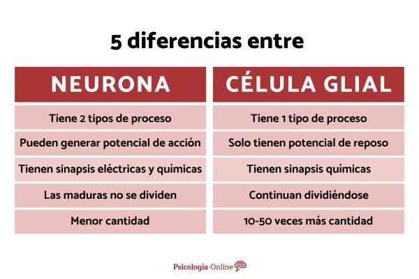
GLIA differs from neurons in several aspects:
- Neurons have two Types of processes; The glia only has one.
- Neurons can generate Action potential: No glial cells, but they have a resting potential.
- Neurons have SINAPSIS that use neurotransmitters; GLIA has chemical synapses.
- Neurons do not continue dividing (not mature, at least); Glial cells yes.
- Another difference between neurons and glial cells is the amount. There are many more glial cells than neurons (at least 10-50 times more).
This article is merely informative, in psychology-online we have no power to make a diagnosis or recommend a treatment. We invite you to go to a psychologist to treat your particular case.
If you want to read more articles similar to Glial cells: what are, types and functions, We recommend that you enter our category of neuropsychology.
- « Prefrontal cortex what is and what functions it does
- Autonomic nervous system what is, parts, functions and characteristics »