

Visual brain Atlas

- 680
- 96
- John Von
Psychoactive offers the reader a visual atlas of the brain with images and explanations of its main components. It is an anatomical atlas through which the most relevant areas at the brain level can be known in depth. Click on the brain area you want.
Visual brain Atlas: parts and functions of the human brain
| " Nervous system | "Hippocampus | " Brainstem |
| "Cerebral tonsil | "Pituitary | "Ventricles |
| "Cerebelle | "Mord | "Somatosensory cortex |
| " Cerebral cortex | "Striated nucleus | "Transversal cuts |
| "DIENTINFALO | " Limbic system | "Front cuts |
| "Septal area |
Nervous system
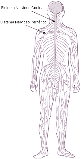
The nervous system is constituted by the organism's nerve tissue and the associated support elements. From a structural or anatomical point of view, the nervous system is divided into two; he Central Nervous System (CNS) and the Peripheral nervous system (SNP). The CNS is formed by the brain and spinal cord, while the SNP comprises specialized nerves, ganglia and receptors.
On the other hand, from the functional point of view the nervous system is divided, into Somatic nervous system and Autonomic nervous system. The somatic system is the part of the nervous system that responds or relates the organism to the external environment, however the autonomous system is in relation to the organic internal environment, performing its own internal regulation and adaptation functions. Both systems do not act independently, but are interrelated and cooperate with each other.
The function of the nervous system consists in receiving the stimuli that come from both the external and internal environment of the organism, organizing this information and making the appropriate response produce.
Stimuli from the external environment are received by the receptors located on the skin, destined to capture general sensations such as pain, touch, pressure and temperature, and by receptors that capture special sensations such as taste, sight, smell, heard, position and movement.
The signals (or impulses) that reach the peripheral nervous system, They are transmitted from these receptors to the central nervous system, where the information is registered and processed. Once registered and processed, the signals are sent from the central nervous system to the different organs in order to provide the appropriate responses.
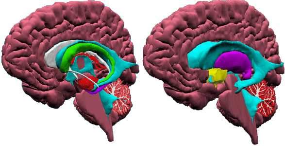
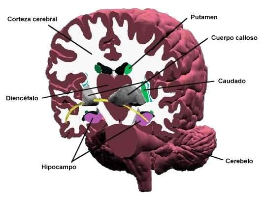
Hippocampus
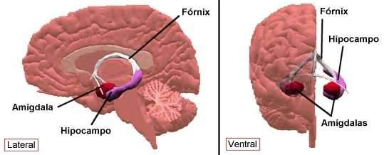
The Hypocampal formation It is located on the average surface of the temporal lobe. Information from the cortex arrives, and in turn sends neuronal signals to the hypothalamus and the septal area through the Fórnix.
The main function of the hippocampus is that of the consolidation of memory and learning. An injury in this area produces amnesia anterograde, that is, the events that occurred after the injury, thus affecting memories of specific facts, but interestingly it does not affect the learning of new capabilities or skills. For example, a person could learn to ride a bicycle after the injury, but I would never remember having seen a bicycle.
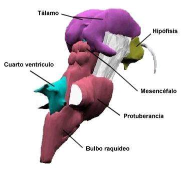
Brainstem
He brainstem It is constituted by the midbrain, the boss and the spinal bulb. All these nervous centers have a similar structure: white substance on the outside with gray substance islets scattered throughout its surface. White substance is made up of nerve fibers that come and go from the brain. He Red nucleus of the midbrain is one of the most prominent gray substance masses. In addition to these rather discrete areas of gray and white substance, the brain stem contains a mixture of both that is called from Reticular formation.
Function
He brainstem It contains numerous reflex centers, the most important of which are the vital centers. These centers are essential for life, since they control respiratory, cardiac and vasomotora activity. In addition to these vital centers, the brain stem contains other centers that control cough, sneezing, hypo, vomiting, suction and swallowing.
Reticular formation exerts two opposite effects on motor activity. On the one hand it facilitates or stimulates such activity, and on the other it depresses it. Studies carried out in the laboratory show that the reticular formation of the brain stem and cerebral adjacent structures (hypothalamus) are necessary for the beginning and maintenance of the state of vigil and consciousness.
The brain trunk contains nuclei corresponding to cranial nerves, and when considering the function of the stem the function of these nerves should not be forgotten. Finally, This is a structure through which the ascending fibers pass from the spinal cord and the descending that are directed to it. Many of these fibers establish connections to different levels with the neurons of the reticular formation and, in some cases, with the neurons of other stem nuclei facilitating the functioning of the reflexes.
Tonsils
The tonsils They are part of the endocrine system, which is formed by a set of glands (thyroid, parathyroid, tonsils, pituitary gland, epiphyses and suparenal gland) that synthesize hormones and release them to the bloodstream. Today it is known that hypothalamus It is responsible for the control of hormonal secretion, and in turn hormones affect the functioning of the nervous system, for this reason the all of the two systems are called Neuroendocrine system.
The endocrine glands control a lot of physiological functions of the organism such as metabolism, homeostasis, growth, reproduction, pain, etc., but They are also involved in human behavior, specifically in emotions, memory, learning or even in pathologies such as depression, anxiety or anorexia nervosa.
Hypophysis
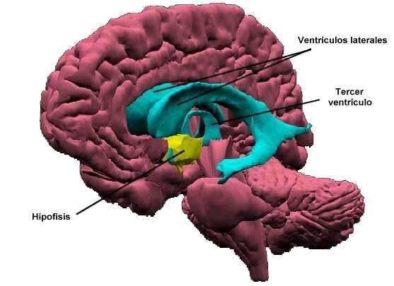
The hypophysis It is located at the base of the brain, together with the hypothalamus and is part of the Neuroendocrine system which is formed by a set of glands (thyroid, parathyroid, tonsils, pituitary gland, epiphyses and suparenal gland) that synthesize hormones and release them to the bloodstream.
The pituitary consists of two parts that work differently: the subsequent pituitary gland either neurohypophysis, which is responsible for storing and releasing hormones synthesized by the hypothalamus (oxytocin and vasopressin). And the Previous pituitary gland either adenohypophysis, that acts as a secretory gland by itself.
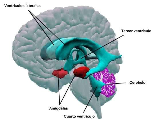
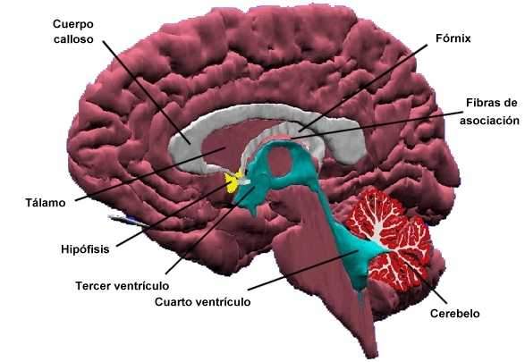
Ventricles
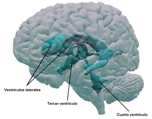
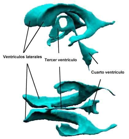
The ventricles cerebrals are composed of several parts: lateral ventricles, the third ventricle and The fourth ventricle. He cerebrospinal fluid It is located inside this ventricular system. He cerebrospinal fluid It is an aqueous liquid that is located in the ventricles and in the subarachnoid spaces. It is produced by the choroid plexus of the ventricles, which are like hairflains covered by epithelial cells. These cells absorb the aqueous fluid of the blood current and secrete inside the ventricles.
The cerebrospinal fluid then passes from the ventricles inside the subarachnoid space through the three openings or holes located in the fourth ventricle. Once in the subarachnoid space, it is absorbed and returns to the bloodstream through the arachnoid membrane, specifically through the arachnoid villi.
Any obstruction in the circulation of cerebrospinal fluid results in the appearance of a ventricular growth known as the name of Hydrocephaly. This condition can cause global head growth if it occurs at an early age, when the bones of the cranial cavity have not been definitively joined. The cerebrospinal fluid, produced continuously from the blood by choroid plexus, cannot be properly reabsorbed in case of hydrocephalus.
The human being has on average a volume of cerebrospinal fluid that ranges around 135 ml. This liquid forms a kind of protective mantle against possible bruises or sudden movements of the head, which otherwise would seriously have an brain integrity. On the other hand, it also serves as a means of referral to the spinal cavity of the liquid volume contained in the cranial cavity. For example, if the cranial cavity penetrates excessive amounts of blood, the derivation of fluid inside the spinal cavity serves to accommodate the additional amounts of blood in the cranial compartment. The brain liquid can also serve for the transport of nutritional substances.
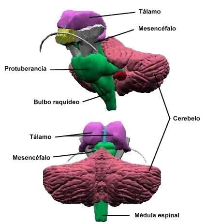
Cerebellum
He cerebellum It is, after the brain, the largest portion of the brain. It occupies the posterior cranial pit and is located under the occipital lobes of the brain, which is separated by a structure called cerebellum store. It consists of two cerebellar hemispheres and an intermediate part called vermis. Binds to the brain stem by three pairs of cerebellar peduncles; These peduncles are fibers that enter and leave the cerebellum, whose surface there are numerous superficial grooves near each other.
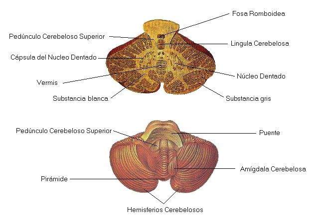
A sagittal cut of the cerebellum shows that outside the cerebellum (in the cerebellar cortex) there is gray substance, and inside the white substance. In the deepest part of the cerebellum are the dentated nuclei. The fourth ventricle occupies a location immediately before the cerebellum.
Microscopic appearance of the cerebellum
The cerebellar cortex is divided into an outer, or molecular layer, and an inner layer, or granulosa. Between the two layers appear cells called Purkinje cells. Although the cells of the two cortical cerebellar layers are small, therefore they are still neurons. Neuroglia is also present.
Cerebellum function
He cerebellum It plays a regulatory role in the coordination of muscle activity, the maintenance of muscle tone and the conservation of balance. The cerebellum requires being constantly informed of what should be done to coordinate muscle activity satisfactorily. To this end, receive information from the different parts of the organism. On the one hand, the cerebral cortex sends a series of fibers that allow cooperation between both structures.
On the other hand, receive information from the muscles and joints, which continuously indicate your position. Finally, receive impulses from the internal ear that keep you informed about the position and movements of the head. The cerebellum requires, then, all this information to be able to carry out the functions that are its own.
To know more about him Cerebelo click here
Spinal cord
The spinal cord is a cylindrical mass of nerve tissue that extends in flow direction from the spinal bulb. The adult's medulla measures approximately 45 cm long and occupies the upper two thirds of the spinal canal. During the early stages of development the spinal cord occupies almost the entire spinal canal, but the rapid growth that the spine experiences immediate. The lower end of the medulla is called a terminal cone.
The spinal cord is divided into 31 segments: 8 cervical, 12 thoracic or dorsal, 5 lumbar, 5 sacros and one coccigeo. The nerves leave the spinal cord along its entire length, in number of one pair for each core segment. The medulla presents two thickening, the cervical and the lumbar. Cervical thickening corresponds to the origin of the nerves that are directed to the upper limb, the lumbar thickening that of nerves that are directed to the lower limb.
Structure
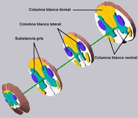
The spinal cord Is made by Gray substance and White substance that adopt a fairly regular distribution. White substance occupies the external part that surrounds the gray substance, and is composed of ascending and descending fibers sustained by neuroglia. When examining a cross section of the medulla, it can. The horizontal part of this H is called gray corner, and each of the tips is called Asta. Consequently, there are two ventral or previous antlers and two dorsal or subsequent antlers.
The white substance is arranged in three columns or fiber cords, anterior or ventral, lateral and posterior or dorsal, which run from one level of the nervous system to another. The fibers that extend from a certain place to another are grouped into beams called fascicles or tracts.
Several cracks run along the spinal cord. In the figure two of these fissures appear, the previous or ventral and the posterior or dorsal. The anterior fissure is deeper and serves to identify the front of the spinal cord.
Function
The gray substance of the spinal cord serves as a reflex center and is part of a distribution center for sensitive and motor tracks.
White substance acts thus from Gran Vía conductor of impulses to the brain and from this.
Somatosensory cortex
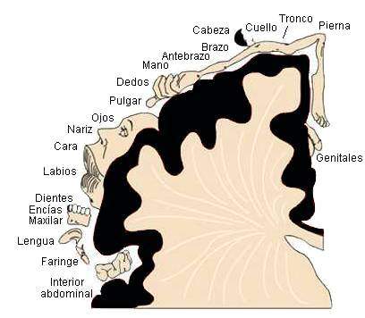
This figure shows the Primary somatosensory areas Of the cerebral cortex, it is a graphic where the areas of the human cortex are represented where the sensations proving from the different parts of the body are recognized, organized and integrate. As can be seen, not all parts the body require the same "quantity" specialized cortex.
The Somesthetic areas o Areas of general sensitivity, are located in the posterior central circumvolution. In this area the sensations of heat, cold, touch, pressure, pain and proprioptíva sensitivity (sense of position and muscle balance) are recorded (sense of position and muscle balance). Each circumvolution receives the sensations from the opposite side of the organism. The arrangement of the body parts represented in the circumvolution also follows an inverse order, so that the sensitive areas of the feet are located at the upper end of the cortex, while the areas for the head occupy the lower end.
The motor areas They are located in the upper central circumvision. Each circumvolution controls the activity of the skeletal muscle that occupies the opposite side of the organism. The various parts of the organism represented in the circumvolution are staggered, from top. Some parts of the organism, such as hand and face, are more represented than others. This is due to the ability of such parties to make more delicate movements.
He Promoting Area, also related to motor activity, occupies a position immediately prior to the precentral circumvolution. The stimulation of this area translates into the appearance of a series of movements of a generalized nature, such as the rotation of the head, trunk turns and general movements of the extremities.
The Language areas, o Broca areas, are located in the frontal lobe. In a right person the areas of language are better developed in the left cerebral cortex. In a left -handed.
The Visual areas They are located in the occipital lobe. In the left occipital lobe, the impulses that originate in the left side of each ocular globe are recorded, while the impulses that originate in the right side are recorded in the right occipital lobe.
The auditory areas They are located in the upper temporal circumvolution. Each temporal lobe receives hearing impulses from both the right and left ear. This is because a considerable number of neurons in charge of transmit.
THE OLFATIVE PRIMARY AREA It is located on the medial surface of the temporal lobe, and the Gusting Primary Area In the anterior face of the posterior central circumvolution of the parietal lobe.
There are other areas called association areas. Those located in the parietal lobe participate in the integration of sensitive information from the somesthetic, auditory, visual and gustatory areas. Parietal association areas correlate information about the various parts of the organism. The associative areas located in the posterior region of the temporal lobe are related to the integration of sensitive data.
Visual and auditory aphasia (inability to understand the oral and written word) can be associated with injuries to these associative areas. The association areas located in the anterior portion of the temporal lobe are related to a variety of experiences, apart from audiovisuals. This previous portion of the temporal lobe has been called psychic cortex because of its relationship with past experiences.
Higher activities such as discernment, reasoning and abstraction also depend on the cerebral cortex. The anterior part of the frontal lobe, called prefrontal area, is in relation to these mental processes characteristic of the human being. The cerebral cortex also exerts an inhibitory influence on the lower parts of the central nervous system.
Cerebral cortex
The cerebral cortex It is the most voluminous part of the brain. A deep cleft, called Longitudinal fissure, divides it into two hemispheres, right and left.
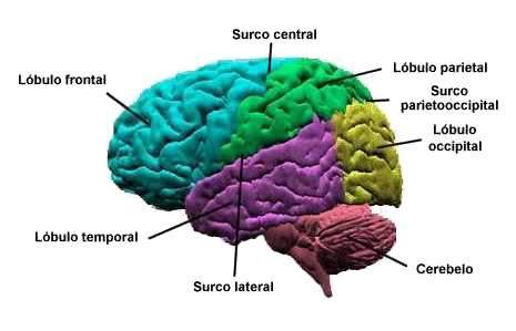
The cerebral cortex It is a fine sheet of interconnected neurons that form a layer of a few millimeters thick and covers the irregular surface of the cerebral hemispheres. The surface of each hemisphere has a set of prominences and furrows (or fissures) that provide the bark a folded appearance, so that only one third of this is exposed to the surface.
Three of these fissures serve to delimit certain areas of the brain.
They are: 1) Central Surco or Rolling Five, 2) side groove or fissure of Silvio, and 3) parietooccipital groove. Eminences between the grooves are called circumvolutions or folds. The anterior central circumvolution is in front of the central groove, and the posterior central circumvolution is placed immediately behind the central groove.
Each hemisphere is divided into four large lobes: frontal, parietal, temporal and occipital. In general, the lobes are located under the bones that bear the same name. Thus, the frontal lobe rests at the depths of the front bone, the parietal lobe under the parietal bone, the temporal lobe under the temporal bone and the occipital lobe under the region corresponding to the protuberance of the occipital.
The aforementioned grooves or fissures act as border structures between some of the brain lobes. The central groove is located between the frontal and parietal lobes. The lateral groove separates the temporal lobe located below the front and parietal lobes located on top. Parietooccipital groove can be visualized on the central surface of the brain.
https: // youtu.BE/IG3XBX0KSM0Striated nucleus
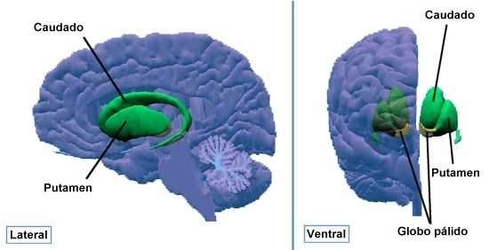
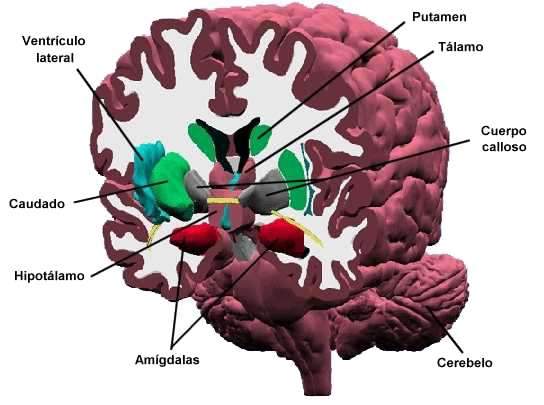
He striated nucleus It is made up: Caudado, putamen and pale balloon. The striated nucleus is inside the cerebral hemispheres, at the base of each hemisphere and its function is related to body movement. This nucleus is part of a higher functional system called Basal Ganglios System, formed by the striated body, the subtálamo and the black substance. The injury of any of these structures can cause alterations in the control of movements (tremor, tics, etc.).
The caudate is cuidos laterally, follows the course of the lateral ventricle. The set of flow and putamen is also called neoestricted, and to the pale balloon Paleoestriate.
Cross cuts
![]()
![]()
![]()
DIENTINFALO
He DIENTINFALO It is a structure located in the central internal part of the cerebral hemispheres. It is located between the hemispheres and the brain trunk, and through it the majority of fibers that are directed towards the cerebral cortex pass.
The diencephalon consists of several parts: Tálamo, Hypothalamus, Subtálamo and Epitálamo.
![]()
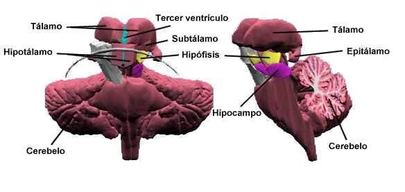
He Tálamo It consists of two ovoid bodies 3 cm long and approximately 1.5 cm thick, which is based on the depth of each cerebral hemisphere. The third ventricle separates each other both attempts, although they remain united thanks to a tasomic tissue bridge called intermediate mass, which extends between the two. Tables are gray substance masses, so they contain neuronal bodies and numerous synaptic connections.
From a functional point of view, the thalamus is a sensitive relief station. Nervous impulses make a talamic level, establishing synapses before continuing their journey to the cerebral cortex. The thalamus also constitutes a primitive sensitive center that serves to record a generalized and imprecise type of sensation.
He hypothalamus It is located, as the name implies, under the thalamus. It presents a wide variety of functions, some of them quite unusual. For example, it produces at least two hormones (oxytocin and vasopressin) and contains centers that regulate the activity of the previous pituitary, the autonomic nervous system, body temperature and water and food intake. In addition, the hypothalamus is related to the state of vigil and emotional sensitivity. In laboratory animals, such as cat, the release of the inhibitory influence that the cerebral cortex exerts on the hypothalamus originates the appearance of bursts of violence before the smallest provocation.
He Subtálamo It is in front of the thalamus and next to the hypothalamus, its main function is related to body movement. The neuronal pathways that cross it go to the thalamus, cerebellum and basal ganglia.
He Epitálamo It is located on the back of the DIENTINFALO, Next to the midbrain. It is formed by the pineal or epiphisi gland and the nuclei of Habénula. Epiphysi is an endocrine gland that secretes melatonin hormone, this secretion is related to the amount of existing sunlight, more light more will be segregated. Habénula has the function of favoring communication between the limbic system and reticular formation.
Limbic system
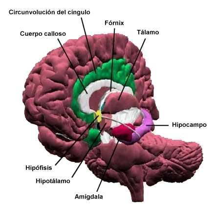
He limbic system It is composed of a set of structures whose function is related to emotional responses, learning and memory. Our personality, our memories and ultimately the fact of being as we are, depends largely on the limbic system.
The components of this system are: amygdala, Tálamo, hypothalamus, hypophysis, hippocampus, The septal area (composed of him Fórnix, Calloso body and association fibers), the orbitofrontal cortex and The Cyngle Circunvolution.
Frontal cuts
Septal area
He septal area It consists of the Calloso Body, the Fórnix and the Association Fibers.
The septal area is closely associated with hippocampus, which is why it also collaborates in the regulation of aggressive behavior, anger and modulation of endocrine activity through the hypothalamus-hypopysarian axis.
In the septal area, sensations such as pleasure and displeasure are generated, as well as eroticism and procreation.